Hamartom Lunge Ct - Sammlung Radiologie Der Lungentumoren / Lung hamartomas are more common in men than in women, and may present additional difficulties in smokers.
Hamartom Lunge Ct - Sammlung Radiologie Der Lungentumoren / Lung hamartomas are more common in men than in women, and may present additional difficulties in smokers.. Authors k ledor, b fish, l chaise, s ledor. Note the low attenuation fat in the mass which is diagnostic of hamartoma. Ct diagnosis of pulmonary hamartomas j comput tomogr. Hamartomas present a peak incidence in the sixth decade of life and are two to four times more common in males than in females. Pulmonary mesenchymal cystic hamartomas (pmhcs) are a rare subtype of pulmonary hamartomas.
2 article feature images from this case Ct diagnosis of pulmonary hamartomas j comput tomogr. Authors k ledor, b fish, l chaise, s ledor. Areas of fat (60%) or calcification (26%) can be seen within the nodule 1. When areas of fat are identified in a nodule on ct.
Pulmonales Hamartom Eref Thieme from eref.thieme.de A lung tumor is an abnormal rate of cell division or cell death in lung tissue or in the airways that lead to the lungs. Case discussion a pulmonary hamartoma is a benign neoplasm composed of normal but disorganized lung tissues, with variable components of cartilage, connective tissue, muscle, fat, and bone. (f) ct image (lung window) obtained 1 year later shows a favorable response to treatment, with marked fibrotic reaction around the healing residual lesions. Hamartomas present a peak incidence in the sixth decade of life and are two to four times more common in males than in females. 2 article feature images from this case Pulmonary (lung) hamartomas are the most common type of benign lung tumor. 1, 2 phs occur mostly as peripheral solitary nodules and. Most common benign tumor of lung, most often with chondromatous element typically solitary, 4 cm, composed of disordered but mature hyaline cartilage, fibrous tissue, smooth muscle, fat, sometimes with calcification / ossification shows some overlapping features with pulmonary chondroma of carney triad, but hamartomas tend to be single rather than multiple, favor men rather than women, and.
Hamartomas present a peak incidence in the sixth decade of life and are two to four times more common in males than in females.
Ct diagnosis of pulmonary hamartomas j comput tomogr. Case discussion a pulmonary hamartoma is a benign neoplasm composed of normal but disorganized lung tissues, with variable components of cartilage, connective tissue, muscle, fat, and bone. Pulmonary hamartoma, the most common benign tumor of the lung, is most often presented as a solitary pulmonary nodule of a peripheral localization. In almost all cases, benign lung tumors require no treatment, but your doctor will probably monitor your tumor for changes. Fat percentages on ct and at histology were compared. They are the most common benign pulmonary neoplasm in adults. Multiple lung hamartomas are rare. Ct criteria for hamartoma included a diameter of 2.5 cm or less, a smooth edge, and focal collections of fat or fat alternating with areas of calcification. When areas of fat are identified in a nodule on ct. Fat is recognized on the ct image in about 60% of cases, and popcornlike calcification or central calcification in about 25% 20 . It is commonly used (either alone or in combination with other chemotherapeutic agents) in the treatment of squamous cell carcinomas (of the head and neck, cervix, and vagina), testicular cancer, and hodgkin lymphoma. Pulmonary (lung) hamartomas are the most common type of benign lung tumor. Hamartoma accounts for 6???8% of solitary lung tumors and 77% of all benign tumors 18, 19 .
Hamartomas present a peak incidence in the sixth decade of life and are two to four times more common in males than in females. If they occur near an airway, they may cause an obstruction resulting in pneumonia and bronchiectasis. Hamartoma is the most common benign tumor of the lung, accounting for 8% of pulmonary neoplasms. They are the most common benign pulmonary neoplasm in adults. Bleomycin is an antitumour antibiotic which was initially isolated from a strain of streptomyces verticillus in 1966.
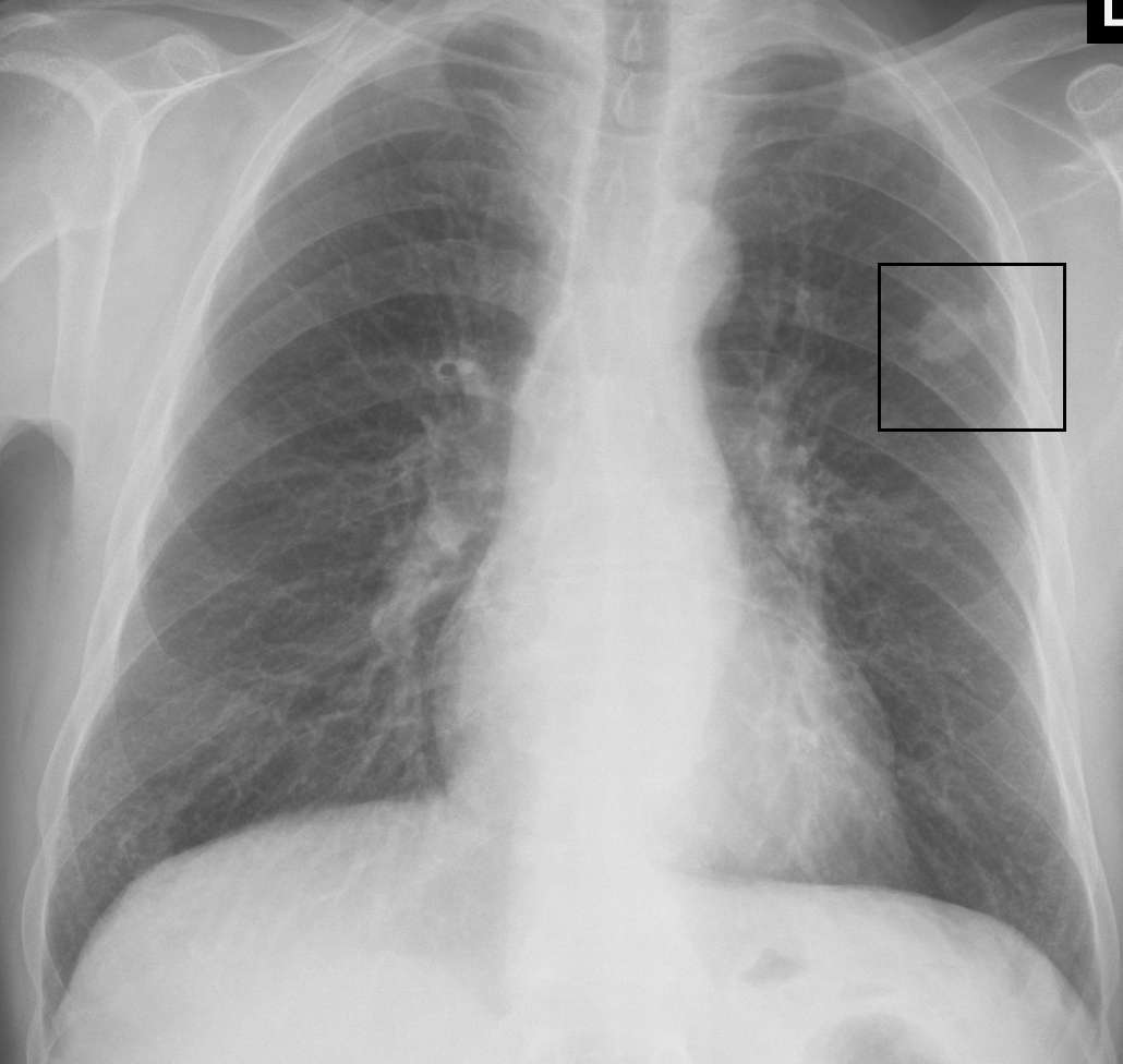
Thorax Iii Nodulare Lungenerkrankungen Dr Adam Domonkos Tarnoki Phd Klinik Fur Radiologie Semmelweis Universitat Pdf Free Download from docplayer.org Hamartoma accounts for 6???8% of solitary lung tumors and 77% of all benign tumors 18, 19 . 2 3 these lesions are usually located in the peripheral parenchyma but have in rare cases. Pulmonary hamartomas can arise in the following lung parts: Ct diagnosis of pulmonary hamartomas j comput tomogr. No case of cancer (n = 283) or metastatic disease (n = 72) fulfilled these criteria. It is commonly used (either alone or in combination with other chemotherapeutic agents) in the treatment of squamous cell carcinomas (of the head and neck, cervix, and vagina), testicular cancer, and hodgkin lymphoma. Endobronchial hamartoma usually appears at ct as a lesion with a smooth edge, focal collections of fat, or fat collections that alternate with foci of calcification. (f) ct image (lung window) obtained 1 year later shows a favorable response to treatment, with marked fibrotic reaction around the healing residual lesions.
Bleomycin is an antitumour antibiotic which was initially isolated from a strain of streptomyces verticillus in 1966.
Ct diagnosis of pulmonary hamartomas j comput tomogr. Imaging of lung hamartomas by multidetector computed tomography and positron emission tomography the present study is the first specifically concerned with the ct and pet/ct characteristics of a pathologically confirmed series of lung hamartomas. Pulmonary mesenchymal cystic hamartomas (pmhcs) are a rare subtype of pulmonary hamartomas. The peak incidence is in the seventh decade of life. Ct criteria for hamartoma included a diameter of 2.5 cm or less, a smooth edge, and focal collections of fat or fat alternating with areas of calcification. Hamartoma is the most common benign tumor of the lung, accounting for 8% of pulmonary neoplasms. The presence of multiple hamartomas in the lungs. Suvmax was correlated with maximum tumor size in pulmonary carcinoids (r = 0.658, p <0.001) and in pulmonary hamartomas (r = 0.672, p <0.001). Pulmonary hamartoma figures 2a and 2b: Types of benign lung tumors include hamartomas, adenomas and papillomas. Lung hamartomas are more common in men than in women, and may present additional difficulties in smokers. Men are affected two to three times more often than women. When areas of fat are identified in a nodule on ct.
Ct diagnosis of pulmonary hamartomas. Pulmonary hamartoma (ph) is the most common benign tumor of the lung and is the third most common cause of solitary pulmonary nodules. The radiological features of pulmonary sclerosing hemangioma (psh) and pulmonary hamartoma are poorly specified. They are the most common benign pulmonary neoplasm in adults. Some of the cavities are thin walled.
Lungenrundherd Wikipedia from upload.wikimedia.org Multiple lung hamartomas are rare. Pulmonary hamartomas can arise in the following lung parts: Pulmonary hamartoma (ph) is the most common benign tumor of the lung and is the third most common cause of solitary pulmonary nodules. Small calcifications are also present. Fat percentages on ct and at histology were compared. Suvmax was correlated with maximum tumor size in pulmonary carcinoids (r = 0.658, p <0.001) and in pulmonary hamartomas (r = 0.672, p <0.001). The area under the receiver operating characteristics curve of suvmax was 0.900 for carcinoids and hamartomas, and 0.722 for typical and atypical carcinoids. Pulmonary (lung) hamartomas are the most common type of benign lung tumor.
Ct diagnosis of pulmonary hamartomas.
Pulmonary hamartomas can arise in the following lung parts: Pulmonary hamartoma, the most common benign tumor of the lung, is most often presented as a solitary pulmonary nodule of a peripheral localization. 2 article feature images from this case Fat percentages on ct and at histology were compared. Men are affected two to three times more often than women. It is commonly used (either alone or in combination with other chemotherapeutic agents) in the treatment of squamous cell carcinomas (of the head and neck, cervix, and vagina), testicular cancer, and hodgkin lymphoma. Hamartomas present a peak incidence in the sixth decade of life and are two to four times more common in males than in females. Visual assessment for macroscopic fat was the most reliable method for diagnosing pulmonary hamartoma. Suvmax was correlated with maximum tumor size in pulmonary carcinoids (r = 0.658, p <0.001) and in pulmonary hamartomas (r = 0.672, p <0.001). Authors k ledor, b fish, l chaise, s ledor. Pulmonary mesenchymal cystic hamartomas (pmhcs) are a rare subtype of pulmonary hamartomas. Hamartoma accounts for 6???8% of solitary lung tumors and 77% of all benign tumors 18, 19 . When areas of fat are identified in a nodule on ct.
Pulmonary (lung) hamartomas are the most common type of benign lung tumor hamar. Pulmonary hamartoma, the most common benign tumor of the lung, is most often presented as a solitary pulmonary nodule of a peripheral localization.


0 Komentar